

The Cleft Palate
Clefts of the palate can occur alone, as an isolated deformity, or in combination with a unilateral or bilateral cleft of the lip. In twenty-five to thirty percent of all individuals with a cleft deformity, the cleft palate is the only cleft problem.
Clefts of the palate may vary considerably. They appear as a single narrow cleft, as a wide cleft which is complete or incomplete, or as a bilateral complete or incomplete cleft in association with a complete or incomplete cleft lip.
Treatment of the child with cleft palate is complex for several reasons. First, the tissue and bony structures of the hard and soft palates are contained within the alveolar arch which means that the only tissue available for closure of the palatal defect must come from the sides of the defect. Second, development of the teeth and alignment of the jaws can be effected. Third, both the hard and soft palates play a vital role in speech development. Fourth, persons with cleft palate often have associated problems with the middle ear.
Classification of Cleft Palate
 Type I a Unilateral with palatal shelves at the same level anteriorly
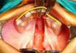 Type I b Unilateral with palatal shelves at the different level anteriorly
 Type II a Bilateral with palatal shelves at the same level anteriorly
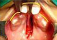 Type II b Bilateral with palatal shelves at the different level anteriorly
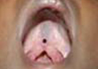 Type IV a Submucons Cleft of Palate
 Type IV b Bifid Uvula
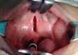 Type III a Isolated Cleft of Soft Palate
 Type III b Isolated Cleft of Hard and Soft Palate
Surgical Repair of the Cleft Palate.
To offer the best chance for normal speech development, we perform the complete repair of the palate in one procedure at the age of twelve months.
With this approach, most patients have normal development of the velopharynx which is essential for normal speech. The Some patients may require future speech therapy and/or and additional operation called pharyngeal flap or pharyngoplasty. The need for additional procedures cannot be determined until the child reaches the age of four to five years and can be adequately assessed for speech development. The surgeical procedure requires about one and a half hours.
The main muscle in the soft palate is called the levator muscle. Normally it forms the soft palate and connects to either side of the throat to help form the velopharynx. It is abnormally attached to the sides of the posterior edge of the hard palate and to the sides to the cleft itself.
The next step in the procedure is to carefully isolate the muscle from their abnormal attachments and to reconstruct the normal muscle sling and configuration in the back of the soft palate
The third and final step of this operation is the complete closure of both nasal and oral layers to create a well-functioning soft palate and to completely close the defect within the hard palate. This type of closure helps to prevent the occurrence of fistulas which are small holes between the oral and nasal cavity within the area of the reconstructed palate. Fistulas result in the leakage of air and fluid into the nasal cavity. The presence of palatal fistulas requires surgical closure since they not only cause leakage, but also hamper normal speech.
 Medial Incisions: uvula to anterior. Lateral incisions: maxilary tuberosity to anterior. Palatopharyngeus muscle and sutured. superficial levator muscles are detached from posterior palate.
 Nasal layer carefully preserved and detached from superior to the palatal shelves and Uvula sutures posteriorly
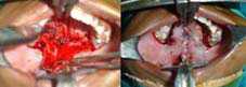 Adaption sutures placed to hold down hard palate mucosa reducing dead space. Sling sutures placed laterally
 Detached muscle sutured to the corresponding muscle on the other side. Palatal mucosa approximated
Postoperative care after cleft palate surgery
Several months are required for complete healing of the reconstructed palate. By four weeks, however, the repair is strong enough to resist damage caused by the baby’s fingers or by eating.
Feeding your child after surgery requires special care. It is imperative to keep your child from sucking during the initial four weeks after surgery. Fluids must be carefully dropped in the mouth from a cup or syringe for the first four weeks after surgery. The child may be offered any fluid that flows form a cup, including pureed food. If the mixture is too thick, it may be diluted with milk. Follow each feeding with water to cleanse the palatal area.
The child will remain in the hospital 24 – 48 hours after surgery. Since the sutures used in palate repair are dissolvable, there is no need to remove them. However, it is important that the child be seen in the hospital for a follow-up visit approximately four weeks after surgery.
